How are auditory stimuli encoded at different
levels of the central auditory pathway?
Greg Detre
Tuesday, 04 December, 2001
Neuro II, essay VI
Dr Andrew King
Introduction
Ohm suggested 100 years ago that the ear and brain deconstruct use Fourier analysis to deconstruct/simplify complex waveforms into the sum of many (simpler) individual sine and cosine waves of appropriate frequencies, phases and amplitudes. Sound is produced by vibrations that result in the alternating compression and rarefaction (increased or decreased pressure) of the surrounding air. The external ear and middle ear form collectively a mechanical transmission system that converts sounds, or air pressure waves, into fluid waves in the inner ear. Vibrations of hair cells are transformed into electrical signals in the auditory nerve
The auditory nerve
Primary auditory afferents � Cell bodies in the spiral ganglion and send their central axons to the pons via the eighth cranial nerve. They fire spontaneously, and increase their firing in response to a tone. Most are sharply tuned.
Frequency coding � two ways:
place coding � for frequencies above 3000Hz, the frequency response of an afferent depends on where along the basilar membrane it is from (tonotopic = frequency-position mapping)
temporal coding � for lower frequencies, afferents fire during a particular phase of the waveform (phase-locking). within the population, each afferent only needs to fire occasionally.
Sound level � auditory afferents respond only to a limited range of sound pressure levels (SPLs). The full range is encoded by populations of afferents with different dynamic ranges. The most sensitive afferents have the highest spontaneous firing rates. Efferents from the superior olivary complex innervate hair cells, reducing their sensitivity, enabling them to respond to high sound levels.
Central auditory pathways � Primary auditory afferents terminate in the cochlear nuclei in the pons. Ventral cochlear axons go to the superior olivary complex on both sides. This projects to the nuclei of the lateral lemniscus, and mainly deals with sound localisation. The dorsal cochlear nucleus projects directly to the contralateral nucleus of the lateral lemniscus. The nuclei of the lateral lemniscus sends axons to the inferior colliculus of the tectum, which projects to the medial geniculate nucleus (MGN). This goes to AI, and is responsible for conscious sound perception. Though the largest auditory pathway is contralateral, extensive connections across the midline ensure interactions between sides.
Cochlear nuclei � Different cell types process different features. Bushy cells signal exact timing information to the (binaural) medial superior olivary nucleus for sound localisation. Stellate cells signal sound level. Many cells are frequency-tuned, further tuned by lateral inhibition.
Tonotopic mapping � Systematic frequency representation (all frequencies are roughly equally represented in humans). In AI, there are isofrequency columns perpendicular to cortical surface, arranged in tonotopic bands.
|
|
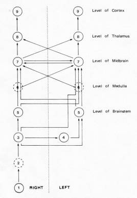

9: Primary
Auditory Cortex
8:� Medial Geniculate Body
7:� Inferior Colliculus
6:� Lateral Lemniscus
5:� Superior Olivary Complex
4:� Trapezoid Body
3:� Cochlear Nucleus
2:� Auditory Nerve Fibers
1:� Cochlear Hair Cells
Most of the fibres in the auditory nerve innervate hair cells
Fibres� tuning curves (the relation between threshold and stimulus frequency) vary, with some having lower thresholds to tones of some frequencies than of others, and each having a �characteristic frequency� (its threshold minimum, ).
Fibres show a sigmoidal relation between firing rate and stimulus intensity.
The frequency resolving power (how selective for a given frequency it is) has been measured by a �quality� factor, calculated as the characteristic frequency divided by the bandwidth of the fibre to tones at an intensity 10 dB abovce the best threshold (known as �Q10�). Fibres with a high Q10 have good frequency selectivity, e.g. the cat�s greatest Q10s are about 8 at about 10kHz.
�During tonal stimulation, auditory nerve fibres fire preferentially during one part of the cycle of the stimulating waveform if the stimulus is below 4-5kHz. The fibres are excited by deflection of the basilar membrane in only one direction.�
�For fibres with characteristic frequencies below 4-5kHz, clicks preferentially evoke responses at certain intervals after the stimulus��
�One tone can reduce, or suppress, the response to another [�two-tone suppression�], even though single tones are only excitatory�. This arises from the non-linear properties of the basilar membrane mechanics, and works for stimuli other than tones too. Also, a stimulus can mask another if it produce a greater firing rate.
The cortex
Romanski et al examined the connectivity of higher auditory cortical areas in macaque monkeys, using a combination of anatomical tracer dyes with electrophysiological recordings. Their results support the ventral/dorsal temporal/parietal what/where processing dichotomy, contributing to functionally distinct regions of the frontal lobe. Further parallelism is evident in the primate auditory cortex, which has three similar primary or primary-like areas, each tonotopically organised and receiving activating inputs directly from the auditory thalamus. The projections from these to 7 or 8 proposed fields seem to provide anatomical support for the beginnings of ventral and dorsal cortical processing streams - they project to largely different portions of the frontal lobe. However, the middle belt area makes connections to the frontal lobe that overlap those of the two putative streams, indicating possible intermediate or additional auditory streams (also analogous to the additional functional streams or 'streams within streams' found in the visual system).
The auditory cortex consists of a:
�core� area � the primary auditory cortex, AI, which receives it input from the main specific auditory relay of the thalamus, the ventral division of the medial geniculate body)
surrounded by a �belt� � receives its input mainly from the other divisions of the medial geniculate
The primary auditory cortex, and some divisions of the belt area, are tonotopically organised. Iso-frequency strips lie at right angles to the line of frequency progression. However, there do not appear to be sudden jumps in frequency as an electrode is moved tangentially in the cortex.
Binaural dominance does seem to be related to the existence of discrete columns (with cells of the same binaural dominance lying in the same radial direction in the cortex, and segregated into discrete strips, running along the cortical surface at roughly right angles to the iso-frequency strips).
Not all neurons in the primary auditory cortex show responses to sound.
There are various shapes of tuning curves in the primary auditory cortex (e.g. broad, multi-peaked), with complex temporal patterns of response.
Many show binaural interactions, suggesting that they code for sound direction. Each cortex predominantly represents sound sources on the contralateral side.
Some cells seem specifically responsive to frequency-modulated stimuli. Othres respond only to complex sounds such as animal calls (though they are not specific detectors for these so much as responding to their basic acoustic elements).
Cortical lesion studies
Initially seemed to show that frequency discrimination is impossible after complete lesions of the auditory cortex. However, frequency discrimination in a series of ongoing pips appears to be possible, while other forms of frequency discrimination are not, apparently purely as a result of task difficulty.
AI seems heavily implicated in sound localisation. Sound locus is coded in a frequency-specific way, such that each iso-frequency strip is involved in coding the source locus for sounds of that frequency.
Upset tasks where the animals have to utilise the temporal dimension of auditory stimuli, or detect/discriminate very short stimuli. The auditory cortex may be necessary for auditory short-term memory, and prolonging the effects of short stimuli.]
The auditory cortex seems to affect he ability to attend to sounds in the contralateral ear.
Function of cortex:
necessary for the analysis of complex sounds
subserves sound localisation and the representation of �auditory space�
necessary for selective attention to auditory stimuli on the basis of source position
serves to inhibit inappropriate motor responses
serves to identify stimuli on an absolute basis
necessary for the discrimination of auditory temporal patterns
necessary for short-term memory when one auditory stimulus has to be related to another later in time
necessary for difficult auditory tasks
Centrifugal pathway
Centrifugal (efferent) auditory pathways parallel the centripetal (afferent) auditory pathways along the entire length of the system, forming a chain which runs from the cortex to the hair cells, often running adjacent to, but not actually within, the tracts and nuclei principally associated with the ascending system.
Electrical activation of the crossed olivocochlear bundle reduces the response of auditory nreve fibres to sound, perhaps because the efferents affect the outer hair cells� signals by affecting the tuning and sensitivity of the mechanical travelling wave on the basilar membrane.
Fibres of the olivocochlear bundle are themselves responsive to sound, and have sharp tuning curves and low thresholds like the afferent fibres. They terminate in corresponding areas of the cochlea , making closed frequency-specific feedback loops. Their activity is also affected by central influences.
Possible functions of the olivocochlear bundle in auditory performance:
may improve the detection of signals in masking noise
may help protect the cochlear from acoustic trauma
may control the mechanical state of the cochlear, compensating for changes in factors like stiffness + static displacemtn foth ebasilar membrance
may be involved in attention
Questions
what�s the difference between sound and all the random motion of the air??? if the answer is �nothing at all�, why don�t we hear a constant cacophony�???
surely because even though the energy of a sound wave is very small, the overall motion of the molecules in the air, like white noise, is not concerted enough to be noticeable
what�s white/pink noise, spectrum???
�random: waveform varies randomly in amplitude over time; amplitude normally distributed around the mean.
white noise: spectrum flat over frequency
pink noise: spectrum energy declines as a function of frequency�
which bits are tonotopic, which aren�t??? how do the tonotopic representations vary???
to what extent does cell type relate to cell function???
which lobe is AI in???
topographic vs anatomic??? correspond to representation and physiology???
how do the various tonotopic representations at the different levels???
what about the descending connections from cortex to thalamus???
Kandel & Schwarz
Pickles
�Single fibres of the auditory nerve are always excited by auditory stimuli and never show sustained inhibition to single stimuli� � eh??? (pg 110)
core/belt = primary/secondary
Instant notes
what/where is the spiral ganglion???
Kaas, Hackett and Tramo
no difference between cochleotopic and tonotopic, is there???
is AII tonotopically organised???